
Tryptofan jako molekularny mikroprzełącznik: integracja analiz statystycznych BioLiP z symulacjami MD receptorów 5-HT₂
DOI:
https://doi.org/10.55225/sti.723Słowa kluczowe:
tryptofan, interakcje białko–ligand, dynamika molekularnaAbstrakt
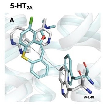
Tryptophan residues play a critical role in protein-ligand recognition owing to their unique aromatic character, high polarizability, and ability to participate in diverse noncovalent interactions, including π-π stacking, cation-π interactions, hydrogen bonding, and hydrophobic contacts. In this study, a two-stage approach was employed to elucidate the functional significance of tryptophan residues within ligand-binding sites across diverse protein systems. First, a large-scale statistical analysis was performed using a curated dataset of protein-ligand complexes extracted from the BioLiP database. The dataset was analysed with respect to enzymatic classification and Gene Ontology enrichment, revealing a pronounced enrichment of tryptophan residues in the binding sites of hydrolases, oxidoreductases, and transferases, as well as strong associations with metal-ion binding, redox-related functions, and membrane or cytoplasmic localization. Within the G protein-coupled receptor (GPCR) superfamily, rhodopsin-like class A receptors were identified as the most prominently represented group featuring tryptophan-mediated ligand interactions. In the second stage, molecular dynamics simulations were conducted for selected serotonin 5-HT₂ receptor subtypes (5-HT₂A, 5-HT₂B, and 5-HT₂C) to provide atomistic insights into the role of the conserved W6.48 residue. The simulations reveal a stable spatial relationship between the ligand and W6.48, characterized by distances consistent with aromatic anchoring, supporting its role in maintaining ligand orientation and binding pocket organization. Collectively, these results highlight tryptophan as a key determinant of ligand recognition and stabilization across both enzymatic and receptor-mediated systems.
Statystyka pobrań
Bibliografia
Bandyopadhyay A, Sarkar R. Site-selective cleavage of peptides and proteins targeting aromatic amino acid residues. RSC Advances. 2025;15(12):9159–9179. https://doi.org/10.1039/d4ra08956a. DOI: https://doi.org/10.1039/D4RA08956A Google Scholar
Barik S. The uniqueness of tryptophan in biology: Properties, metabolism, interactions and localization in proteins. International Journal of Molecular Sciences. 2020;21(22):8776. https://doi.org/10.3390/ijms21228776. DOI: https://doi.org/10.3390/ijms21228776 Google Scholar
Calinsky R, Levy Y. Aromatic residues in proteins: Re-evaluating the geometry and energetics of π-π, cation−π, and CH−π interactions. Journal of Physical Chemistry B. 2024;128(36):8687–8700. https://doi.org/10.1021/acs.jpcb.4c04774. DOI: https://doi.org/10.1021/acs.jpcb.4c04774 Google Scholar
Dougherty DA. The cation-π interaction. Accounts of Chemical Research. 2013;46(4):885-893. https://doi.org/10.1021/ar300265y. DOI: https://doi.org/10.1021/ar300265y Google Scholar
Szczygieł M, Derewenda U, Scheiner S, Minor W, Derewenda ZS. A structural role for tryptophan in proteins, and the ubiquitous TRP Cδ1-H···O=C (backbone) hydrogen bond. Acta Crystallographica Section D Structural Biology. 2024;80(Pt 7):551–562. https://doi.org/10.1107/S2059798324005515. DOI: https://doi.org/10.1107/S2059798324005515 Google Scholar
Brylinski M. Aromatic interactions at the ligand-protein interface: Implications for the development of docking scoring functions. Chemical Biology and Drug Design. 2018;91(2):380–390. https://doi.org/10.1111/cbdd.13084. DOI: https://doi.org/10.1111/cbdd.13084 Google Scholar
Shao J, Kuiper BP, Thunnissen AMWH, Cool RH, Zhou L, Huang C, Liu J, et al. The role of tryptophan in π interactions in proteins: An experimental approach. Journal of the American Chemical Society. 2022;144(30):13815– Google Scholar
–13822. https://doi.org/10.1021/jacs.2c04986. DOI: https://doi.org/10.1021/jacs.2c04986 Google Scholar
Lopez-Balastegui M, Stepniewski TM, Kogut-Günthel MM, Di Pizio A, Rosenkilde MM, Mao J, Selent J. Relevance of G protein-coupled receptor (GPCR) dynamics for receptor activation, signalling bias and allosteric modulation. British Journal of Pharmacology. 2025;182(14):3211–3224. https://doi.org/10.1111/bph.16495. DOI: https://doi.org/10.1111/bph.16495 Google Scholar
Latorraca NR, Venkatakrishnan AJ, Dror RO. GPCR dynamics: Structures in motion. Chemical Reviews. 2017;117(1):139–155. https://doi.org/10.1021/acs.chemrev. DOI: https://doi.org/10.1021/acs.chemrev.6b00177 Google Scholar
b00177. Google Scholar
Trzaskowski B, Latek D, Yuan S, Ghoshdastider U, Debinski A, Filipek S. Action of molecular switches in GPCRs: Theoretical and experimental studies. Current Medicinal Chemistry. 2012;19(8):1090–1109. https://doi.org/10.2174/092986712799320556 DOI: https://doi.org/10.2174/092986712799320556 Google Scholar
Plazinska A, Plazinski W, Luchowski R, Wnorowski A, Grudzinski W, Gruszecki WI. Ligand-induced action of the W286⁶.⁴⁸ rotamer toggle switch in the β₂-adrenergic receptor. Physical Chemistry Chemical Physics 2017;20(1):581–594. https://doi.org/10.1039/c7cp04808d. DOI: https://doi.org/10.1039/C7CP04808D Google Scholar
Nichols DE, Nichols CD. Serotonin receptors. Chemical Reviews. 2008;108(5):1614–1641. https://doi.org/10.1021/cr078224o. DOI: https://doi.org/10.1021/cr078224o Google Scholar
Voitel M, Pankonin M, Vogel A, Leitner K, Kaiser A, Huster D, Hildebran PW, Smith AA. Fine-tuning of the neuropeptide Y1 G protein-coupled receptor by the tryptophan⁶.⁴⁸ “Toggle Switch.” Journal of the American Chemical Society. 2026;148(1):194–205. https://doi.org/10.1021/jacs.5c07143. DOI: https://doi.org/10.1021/jacs.5c07143 Google Scholar
Lee Y, Basith S, Choi S. Recent advances in structure-based drug design targeting class A G protein-coupled receptors utilizing crystal structures and computational simulations. Journal of Medicinal Chemistry. 2018;61(1):1–46. https://doi.org/10.1021/acs.jmedchem.6b01453. DOI: https://doi.org/10.1021/acs.jmedchem.6b01453 Google Scholar
Kim J, Kim J, Choi C, Bae J, Choi HJ. Structural insights into GPCR signalling activated by peptide ligands: From molecular mechanism to therapeutic application. Experimental & Molecular Medicine. 2025;57(7):1467–1481. https://doi.org/10.1038/s12276-025-01497-y. DOI: https://doi.org/10.1038/s12276-025-01497-y Google Scholar
Yuan S, Hu Z, Filipek S, Vogel H. W246⁶.⁴⁸ opens a gate for a continuous intrinsic water pathway during activation of the adenosine A₂A receptor. Angewandte Chemie: International Edition. 2015;54(2):556–559. https://doi.org/10.1002/anie.201409679. DOI: https://doi.org/10.1002/anie.201409679 Google Scholar
Peng Y, McCorvy JD, Harpsøe K, Lansu K, Yuan S, Popov P, Qu L, Pu M, Che T, Nikolajsen LF, Huang XP, Wu Y, Shen L, Bjørn-Yoshimoto WE, Ding K, Wacker D, Han GW, Cheng J, Katritch V, Jensen AA, Liu ZJ. 5-HT2C receptor structures reveal the structural basis of GPCR polypharmacology. Cell. 2018;72(4):719–730.e14. https://doi.org/10.1016/j.cell.2018.01.001. DOI: https://doi.org/10.1016/j.cell.2018.01.001 Google Scholar
Yang J, Roy A, Zhang Y. BioLiP: A semi-manually curated database for biologically relevant ligand-protein interactions. Nucleic Acids Research. 2013;41:D1096-1103. https://doi.org/10.1093/nar/gks966. DOI: https://doi.org/10.1093/nar/gks966 Google Scholar
Zhang C, Zhang X, Freddolino PL, Zhang Y. BioLiP2: an updated structure database for biologically relevant ligand-protein interactions. Nucleic Acids Research. 2024.52(D1):D404–412. https://doi.org/10.1093/nar/gkad630. DOI: https://doi.org/10.1093/nar/gkad630 Google Scholar
Lee J, Cheng X, Jo S, MacKerell AD, Klauda JB, Im W. CHARMM-GUI Input Generator for NAMD, Gromacs, Amber, Openmm, and CHARMM/OpenMM simulations using the CHARMM36 Additive Force Field. Biophysical Journal. 2016;110(3 suppl. 1):641a. https://doi.org/10.1016/j.bpj.2015.11.3431 DOI: https://doi.org/10.1016/j.bpj.2015.11.3431 Google Scholar
Kern NR, Lee J, Choi YK, Im W. CHARMM-GUI Multicomponent Assembler for modeling and simulation of complex multicomponent systems. Nature Communications. 2024;15(1):5459. https://doi.org/10.1038/s41467-024-49700-4. DOI: https://doi.org/10.1038/s41467-024-49700-4 Google Scholar
Lee J, Patel DS, Ståhle J, Park S-J, Kern NR, Kim S,Lee J, Cheng X, Valvano MV, Holst O, Knirel Y, Qi Y, Jo S, Klauda JB, Widmalm G, Im W. CHARMM-GUI Membrane Builder for complex biological membrane simulations with glycolipids and lipoglycans. Journal of Chemical Theory and Computation. 2019;15(1):775–786. https://doi.org/10.1021/acs.jctc.8b01066. DOI: https://doi.org/10.1021/acs.jctc.8b01066 Google Scholar
Pytliak M, Vargová V, Mechírová V, Felšöci M. Serotonin receptors—from molecular biology to clinical applications. Physiological Research. 2011;60(1):15–25. https://doi.org/10.33549/physiolres.931903. DOI: https://doi.org/10.33549/physiolres.931903 Google Scholar
Manglik A, Kruse AC. Structural basis for G protein-coupled receptor activation. Biochemistry. 2017;56(42):5628––5634. https://doi.org/10.1021/acs.biochem.7b00747. DOI: https://doi.org/10.1021/acs.biochem.7b00747 Google Scholar
Weis WI, Kobilka BK. The molecular basis of G protein-coupled receptor activation. Annual Review of Biochemistry. 2018;87:897–919. https://doi.org/10.1146/annurev-biochem-060614-033910. DOI: https://doi.org/10.1146/annurev-biochem-060614-033910 Google Scholar
Pándy-Szekeres G, Munk C, Tsonkov TM, Mordalski S, Harpsøe K, Hauser AS, Bojarski AJ, Gloriam DE. GPCRdb in 2018: Adding GPCR structure models and ligands. Nucleic Acids Research. 2018;46(D1):D440–446. https://doi.org/10.1093/nar/gkx1109. DOI: https://doi.org/10.1093/nar/gkx1109 Google Scholar
Pobrania
Opublikowane
Jak cytować
Numer
Dział
Licencja
Prawa autorskie (c) 2026 Karina Pakosz, Paweł Śliwa

Utwór dostępny jest na licencji Creative Commons Uznanie autorstwa – Na tych samych warunkach 4.0 Miedzynarodowe.