Application of Fragment Molecular Orbital Method to investigate dopamine receptors
DOI:
https://doi.org/10.5604/01.3001.0013.5526Keywords:
fragment molecular orbital, molecular dynamic, dopamine receptorsAbstract
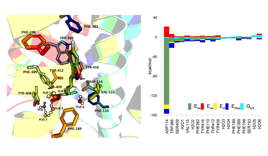
GPCRs are a vast family of seven-domain transmembrane proteins. This family includes dopamine receptors (D1, D2, D3, D4, and D5), which mediate the variety of dopamine-controlled physiological functions in the brain and periphery. Ligands of dopamine receptors are used for managing several neuropsychiatric disorders, including bipolar disorder, schizophrenia, anxiety, and Parkinson’s disease. Recent studies have revealed that dopamine receptors could be part of multiple signaling cascades, rather than of a single signaling pathway. For these targets, a variety of experimental and computational drug design techniques are utilized. In this work, dopamine receptors D2, D3, and D4 were investigated using molecular dynamic method as well as computational ab initio Fragment Molecular Orbital method (FMO), which can reveal atomistic details about ligand binding. The results provided useful insights into the significances of amino acid residues in ligand binding sites. Moreover, similarities and differences between active-sites of three studied types of receptors were examined.
Downloads
References
Beaulieu JM, Gainetdinov RR. The Physiology, Signaling, and Pharmacology of Dopamine Receptors. Pharmacol Rev. 2011;63:182–217. Google Scholar
Vallone D, Picetti R, Borrelli E. Structure and function of dopamine receptors. Neurosci Biobehav Rev. 2000;24(1):125–32. Google Scholar
Beaulieu JM, Gainetdinov RR, Caron MG. The Akt-GSK-3 signaling cascade in the actions of dopamine. Trends Pharmacol Sci. 2007;28(4):166–72. Google Scholar
Wang M, Wong AH, Liu F. Interactions between NMDA and dopamine receptors: A potential therapeutic target. Brain Res. 2012;1476:154–63. Google Scholar
Damian M, Pons V, Renault P, M’Kadmi C, Delort B, Hartmann L, et al. GHSR-D2R heteromerization modulates dopamine signaling through an effect on G protein conformation. Proc Natl Acad Sci. 2018;115(17):4501–6. Google Scholar
Dong N, Lee DWK, Sun HS, Feng ZP. Dopamine-mediated calcium channel regulation in synaptic suppression in l. Stagnalis interneurons. Channels. 2018;12(1):153–73. Google Scholar
Hasbi A, Fan T, Alijaniaram M, Nguyen T, Perreault M, O’Dowd BF, et al. Calcium signaling cascade links dopamine D1–D2 receptor heteromer to striatal BDNF production and neuronal growth. Proc Natl Acad Sci. 2009;106(50):21477–21382. Google Scholar
Hasbi A, O’Dowd BF, George SR. Heteromerization of dopamine D2 receptors with dopamine D1 or D5 receptors generates intracellular calcium signaling by different mechanisms. Curr Opin Pharmacol. 2010;10(1):93–9. Google Scholar
Iwakura Y, Nawa H, Sora I, Chao M V. Dopamine D1 receptor-induced signaling through TrkB receptors in striatal neurons. J Biol Chem. 2008;283(23):15799–806. Google Scholar
Kotecha SA, Oak JN, Jackson MF, Perez Y, Orser BA, Van Tol HHM, et al. A D2 class dopamine receptor transactivates a receptor tyrosine kinase to inhibit NMDA receptor transmission. Neuron. 2002;35(6):1111–22. Google Scholar
Marion S, Urs NM, Peterson SM, Sotnikova TD, Beaulieu J-M, Gainetdinov RR, et al. Dopamine D2 Receptor Relies upon PPM/PP2C Protein Phosphatases to Dephosphorylate Huntingtin Protein. J Biol Chem. 2014;289(17):11715–24. Google Scholar
Medvedev IO, Ramsey AJ, Masoud ST, Bermejo MK, Urs N, Sotnikova TD, et al. D 1 dopamine receptor coupling to PLCβ regulates forward locomotion in mice. J Neurosci. 2013;33(46):18125–33. Google Scholar
Luderman KD, Conroy JL, Free RB, Southall N, Ferrer M, Sanchez-Soto M, et al. Identification of positive allosteric modulators of the D 1 dopamine receptor that act at diverse binding sites S. Mol Pharmacol. 2018;94(4):1197–209. Google Scholar
Shen Y, McCorvy JD, Martini ML, Rodriguiz RM, Pogorelov VM, Ward KM, et al. D2 Dopamine Receptor G Protein-Biased Partial Agonists Based on Cariprazine. J Med Chem. 2019;62(9):4755–71. Google Scholar
Bonifazi A, Yano H, Guerrero AM, Kumar V, Hoffman AF, Lupica CR, et al. Novel and Potent Dopamine D 2 Receptor Go-Protein Biased Agonists . ACS Pharmacol Transl Sci. 2019;2(1):52–65. Google Scholar
Chun LS, Vekariya RH, Free RB, Li Y, Lin DT, Su P, et al. Structure-activity investigation of a G protein-biased agonist reveals molecular determinants for biased signaling of the D 2 dopamine receptor. Front Synaptic Neurosci. 2018;10:1–18. Google Scholar
Gordon MS, Fedorov DG, Pruitt SR, Slipchenko L V. Fragmentation methods: A route to accurate calculations on large systems. Chem Rev. 2012;112(1):632–72. Google Scholar
Fedorov DG. The fragment molecular orbital method: theoretical development, implementation in GAMESS, and applications. Wiley Interdiscip Rev Comput Mol Sci. 2017;7(6):1–17. Google Scholar
Fedorov DG, Avramov P V., Jensen JH, Kitaura K. Analytic gradient for the adaptive frozen orbital bond detachment in the fragment molecular orbital method. Chem Phys Lett. 2009;477(1–3):169–75. Google Scholar
Fedorov DG, Jensen JH, Deka RC, Kitaura K. Covalent bond fragmentation suitable to describe solids in the fragment molecular orbital method. J Phys Chem A. 2008;112(46):11808–16. Google Scholar
Fedorov DG, Kitaura K. The importance of three-body terms in the fragment molecular orbital method. J Chem Phys. 2004;120:6832–40. Google Scholar
Fedorov DG, Kitaura K. Second order Møller-Plesset perturbation theory based upon the fragment molecular orbital method. J Chem Phys. 2004;121(6):2483–90. Google Scholar
Shimamura K, Ishimura H, Kobayashi I, Kadoya R, Kurita N, Kawai K, et al. Molecular dynamics and ab initio FMO calculations on the effect of water molecules on the interactions between androgen receptor and its ligand and cofactor. 4th IGNITE Conf 2016 Int Conf Adv Informatics Concepts, Theory Appl ICAICTA 2016. 2016;1–6. Google Scholar
Fedorov DG, Kitaura K. Pair interaction energy decomposition analysis. J Comput Chem. 2007;28(1):222–37. Google Scholar
Chudyk EI, Sarrat L, Aldeghi M, Fedorov DG, Bodkin MJ, James T, et al. Exploring GPCR-ligand interactions with the fragment molecular orbital (FMO) method. Methods Mol Biol. 2018;1705:179–95. Google Scholar
Akimov A V. Nonadiabatic Molecular Dynamics with Tight-Binding Fragment Molecular Orbitals. J Chem Theory Comput. 2016;12(12):5719–36. Google Scholar
Doi H, Okuwaki K, Mochizuki Y, Mochizuki Y, Ozawa T, Yasuoka K. Dissipative particle dynamics (DPD) simulations with fragment molecular orbital (FMO) based effective parameters for 1-Palmitoyl-2-oleoyl phosphatidyl choline (POPC) membrane. Chem Phys Lett. 2017;684:427–32. Google Scholar
Gaus M, Cui Q, Elstner M. Density functional tight binding: Application to organic and biological molecules. Wiley Interdiscip Rev Comput Mol Sci. 2014;4(1):49–61. Google Scholar
Ishimura H, Tomioka S, Kadoya R, Shimamura K, Okamoto A, Shulga S, et al. Specific interactions between amyloid-Β peptides in an amyloid-Β hexamer with three-fold symmetry: Ab initio fragment molecular orbital calculations in water. Chem Phys Lett. 2017;672:13–20. Google Scholar
Kobayashi I, Takeda R, Suzuki R, Shimamura K, Ishimura H, Kadoya R, et al. Specific interactions between androgen receptor and its ligand: ab initio molecular orbital calculations in water. J Mol Graph Model. 2017;75:383–9. Google Scholar
Komeiji Y, Okiyama Y, Mochizuki Y, Fukuzawa K. Interaction between a Single-Stranded DNA and a Binding Protein Viewed by the Fragment Molecular Orbital Method. Bull Chem Soc Jpn. 2018;91(11):1596–605. Google Scholar
Ozawa M, Ozawa T, Ueda K. Application of the fragment molecular orbital method analysis to fragment-based drug discovery of BET (bromodomain and extra-terminal proteins) inhibitors. J Mol Graph Model. 2017;74:73–82. Google Scholar
Sawada T, Hashimoto T, Nakano H, Suzuki T, Ishida H, Kiso M. Why does avian influenza A virus hemagglutinin bind to avian receptor stronger than to human receptor? Ab initio fragment molecular orbital studies. Biochem Biophys Res Commun. 2006;351(1):40–3. Google Scholar
Simoncini D, Nakata H, Ogata K, Nakamura S, Zhang KYJ. Quality Assessment of Predicted Protein Models Using Energies Calculated by the Fragment Molecular Orbital Method. Mol Inform. 2015;34(2–3):97–104. Google Scholar
Śliwa P, Kurczab R, Bojarski AJ. ONIOM and FMO-EDA study of metabotropic glutamate receptor 1: Quantum insights into the allosteric binding site. Int J Quantum Chem. 2018;118(15):e25617. Google Scholar
Śliwa P, Kurczab R, Kafel R, Drabczyk A, Jaśkowska J. Recognition of repulsive and attractive regions of selected serotonin receptor binding site using FMO-EDA approach. J Mol Model. 2019 6;25(5):114. Google Scholar
Steinmann C, Ibsen MW, Hansen AS, Jensen JH. FragIt: A Tool to Prepare Input Files for Fragment Based Quantum Chemical Calculations. PLoS One. 2012;7(9). Google Scholar
Takeda R, Kobayashi I, Suzuki R, Kawai K, Kittaka A, Takimoto-Kamimura M, et al. Proposal of potent inhibitors for vitamin-D receptor based on ab initio fragment molecular orbital calculations. J Mol Graph Model. 2018;80:320–6. Google Scholar
Terauchi Y, Suzuki R, Takeda R, Kobayashi I, Kittaka A, Takimoto-Kamimura M, et al. Ligand chirality can affect histidine protonation of vitamin-D receptor: ab initio molecular orbital calculations in water. J Steroid Biochem Mol Biol. 2019;186:89–95. Google Scholar
Vuong VQ, Nishimoto Y, Fedorov DG, Sumpter BG, Niehaus TA, Irle S. The Fragment Molecular Orbital Method Based on Long-Range Corrected Density-Functional Tight-Binding. J Chem Theory Comput. 2019;15(5):3008–20. Google Scholar
Yoshino R, Yasuo N, Inaoka DK, Hagiwara Y, Ohno K, Orita M, et al. Pharmacophore modeling for anti-Chagas drug design using the fragment molecular orbital method. PLoS One. 2015;10(5):1–15. Google Scholar
Willighagen EL, Waagmeester A, Spjuth O, Ansell P, Williams AJ, Tkachenko V, et al. The ChEMBL database as linked open data. J Cheminform. 2013;5(5):1–12. Google Scholar
Phillips JC, Braun R, Wang W, Gumbart J, Tajkhorshid E, Villa E, et al. Scalable molecular dynamics with NAMD. J Comput Chem. 2005;26(16):1781–802. Google Scholar
Vonommeslaeghe K, Hatcher E, Acharya C, Kundu S, Zhong S, Shim J, et al. CHARMM General Force Field: A Force Field for Drug-Like Molecules Compatible with the CHARMM All-Atom Additive Biological Force Fields. J Comput Chem. 2009;31(4):671–90. Google Scholar
Lomize MA, Pogozheva ID, Joo H, Mosberg HI, Lomize AL. OPM database and PPM web server: resources for positioning of proteins in membranes. Nucleic Acids Res. 2012;40(D1):D370–6. Google Scholar
Pettersen EF, Goddard TD, Huang CC, Couch GS, Greenblatt DM, Meng EC, et al. UCSF Chimera - A visualization system for exploratory research and analysis. J Comput Chem. 2004;25(13):1605–12. Google Scholar
Suenaga M. FACIO. Department of Chemistry, Graduate School of Sciences, Kyushu University, Japan. Google Scholar
Fedorov DG, Kitaura K, Li H, Jensen JH, Gordon MS. The polarizable continuum model (PCM) interfaced with the fragment molecular orbital method (FMO). J Comput Chem. 2006 Jun;27(8):976–85. Google Scholar
Ballesteros JA, Weinstein H. Integrated Methods for the Construction of Three-Dimensional Models and Computational Probing of Structure-Function Relations in G Protein-Coupled Receptors. Methods Neurosci. 1995;25:366–428. Google Scholar
Kurek J, Kwaśniewska P, Myszkowski K, Cofta G. Antifungal , anticancer and docking studies of colchiceine complexes with monovalent metal cation salts. Chem Biol Drug Des. 2019;00:1–14. Google Scholar
Kurczab R, Śliwa P, Rataj K, Kafel R, Bojarski AJ. Salt Bridge in Ligand–Protein Complexes—Systematic Theoretical and Statistical Investigations. J Chem Inf Model. 2018;58(11):2224–38. Google Scholar

Downloads
Published
How to Cite
Issue
Section
License
Copyright (c) 2019 University of Applied Sciences in Tarnow, Poland & Authors

This work is licensed under a Creative Commons Attribution-NonCommercial 4.0 International License.


